Featured

HYPOXIA

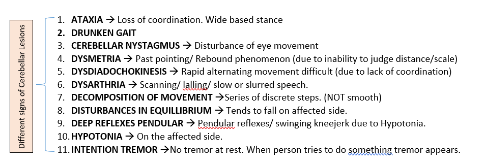
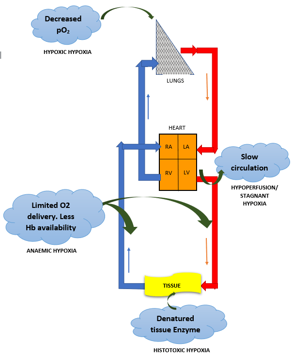
Hypoxia is O2 deficiency at the tissue level. Traditionally, hypoxia has been divided into four types.
Numerous other classifications have been used, but the four type system is the one which is most widely used and excepted:

TYPES OF HYPOXIA

HYPOXIC HYPOXIA

Most common type. A problem seen in NORMAL individuals at HIGH ALTITUDE, complication of PNEUMONIA and many RESPIRATORY COMPLICATIONS. It is a is a condition of reduced arterial pO2

Low arterial pO2

Arterio-venous pO2 difference – LOW

Ventilation – perfusion Imbalance

Venous to arterial Shunt

TREATMENT -Treat the cause. OXYGEN THERAPY

HYPOXIC HYPOXIA

ANAEMIC HYPOXIA

limited ability to deliver O2 to the active tissues. In anemia hypoxia is seen during exercise. Carbon monoxide poisoning – carboxyhemoglobin/COHb, is formed which shifts available HbO2 curve to left and CO has 210 times affinity to Hb than O2 which makes release of CO very slow.

Arterial pO2 normal

Arterio-venous pO2 difference – NORMAL

TREATMENT – Hyperbaric OXYGEN therapy

STAGNANT HYPOXIA

Due to slow circulation and is a problem in organs such as the kidneys and heart OR during shock. The liver and possibly the brain are damaged by hypo perfusion during CONGESTIVE HEART FAILURE.

↑  pCO2 at tissue.

Arterio-venous pO2 difference – INCREASED

TREATMENT – Treat the underlying cause.

HISTOTOXIC HYPOXIA

Due to inhibition of tissue oxidative processes. CAUSE – cyanide poisoning. Cyanide inhibits cytochrome oxidase and possibly other enzymes at TISSUE level. O2 utilization is inhibited.

Tissue cant utilize pO2

Arterio-venous pO2 difference – ABSENT/ALMOST NIL

TREATMENT – Methylene blue or nitrites are used to treat cyanide poisoning. They act by forming methemoglobin, which then reacts with cyanide to form cyanmethemoglobin, a nontoxic compound.

HYPERBARIC Oxygen

Different types of HYPOXIA

Administration of oxygen-rich gas mixtures is of very limited value in hypoperfusion/Stagnant, anemic, and histotoxic hypoxia. Hyperbaric Oxygen helps by increasing the fraction of DISSOLVED Oxygen. Except SHUNT, in other forms of hypoxic hypoxia, O2 is of great benefit.

EFFECT OF HYPOXIA

Effect of Hypoxia

HAPPY HYPOXIA/HYPOXAEMIA IN COVID-19

COVID -19 infection can bring down the Oxygen saturation, as low as 40 %.

Hypoxia is a warning signal for imminent failure of vital body organs like the kidneys, brain, heart and is usually accompanied by prominent breathlessness. Many patients with pronounced arterial hypoxemia don’t even experience a sense of dyspnea. As a result, in the initial stages of sickness, the COVID-19 patient, on the outside, appears to be alright and “happy” due to lack of any external signals.

The trick to life is to just keep breathing. – Johnny Lung

Featured

THALAMUS

Thalamus in Greek means “Ante chamber” or “Inner chamber“. It was discovered way back in the 2nd Century AD, when Galen traced the optic-nerve fibers to an oval mass closely associated with the ventricles.

It is considered as head ganglion of all sensory systems.It integrates & processes all sensory impulses. All sensory pathways (EXCEPTION – Olfactory pathway) that proceeds to the cortex first relays in the thalamus before reaching its destination.

THALAMIC NUCLEI

NUCEI OF THALAMUS

FUNCTIONAL CLASSIFICATION

NON-SPECIFIC N– RECEIVE IMPULSES FOR DIFFUSE SECONDARY RESPONSES FROM THE RETICULAR ACTIVATING SYSTEM & PROJECT DIFFUSELY TO NEOCORTEX. SPECIFIC NUCLEI – RECEIVE SPECIFIC SENSATION & PROJECT TO SPECIFIC PORTION OF NEOCORTEX & LIMBIC SYSTEM
DEPENDING UPON THE TYPE OF SENSATION, THE SPECIFIC PROJECTION NUCLEI CAN BE DIVIDED INTO 4 GROUPS

CONNECTIONS OF THALAMUS

Afferent & efferent connections of various thalamic nuclei is based on the functional classification:

SPECIFIC NUCLEI OF THALAMUS

  1. PRINCIPAL RELAY
PRINCIPAL RELAY

VENTRAL POSTERIOR NUCLEUS (VPN) It has 2 divisions : VPL( Ventro posterior Lateral) & VPM (Ventro Postero Medial) {sensory relay Nuclei}

MEDIAL GENICULATE BODY – Receive topically organised projection of auditory fibers. {sensory relay Nuclei}

LATERAL GENICULATE BODY – Show an orderly organized representation of the retina {sensory relay Nuclei} AFFERENT: OPTIC TRACT, SUPERIOR COLLICULUS EFFERENT : VISUAL CORTEX (17,18,19) FUNCTION : VISION

VENTRAL LATERAL NUCLEUS (VLN) : Chief motor nucleus of Thalamus . Acts as a relay centre for cerebellar impulses. {Motor Control Nuclei}

VLN OF THALAMUS

VENTRAL ANTERIOR NUCLEUS : Involved in the programming of movements controlled by the basal Ganglia. {Motor Control Nuclei}

VENTRAL ANTERIOR NUCLEUS

ANTERIOR NUCLEUS : Belongs to Papez Circuit of Limbic system. Concerned with recent memory & emotions {Visceral Efferent Control Nuclei}

ANTERIOR NUCLEUS OF THALAMUS

2. ASSOCIATION NUCLEUS

LATERAL NUCLEUS : AFFERENT : Parietal lobe of cortex, Sup colliculus, principal relay N. EFFERENT : Parietal association area (language), Cingulate Gyrus. FUNCTION : Language comprehension, speech, other integrated function

DORSOMEDIAL NUCLEUS: AFFERENT : Relay N, Hypothalamus, Amygdala, Pre-frontal Cortex, Olfactory information. EEFERENT : Pre-frontal Cortex (area- 8,9,10,11), Newer olfactory pathway to orbito-frontal cortex. FUNCTION : Processing of Crude somatic, visceral sensation, perception of olfaction, elaboration of thought, word formation.

PULVINAR : AFFERENT : Relay N, Parietal, Temporal, Occipital Cortex, Sup Colliculus. EFFERENT : Visual association area (lateral part) & Auditory association area (medial part) FUNCTION : Integration of visual, auditory & Somasthetic information.

NON-SPECIFIC THALAMIC NUCLEI

NON-SPECIFIC THALAMIC NUCLEI

FUNCTIONS OF THALAMUS

FUNCTIONS OF THALAMUS

THALAMIC SYNDROME

Dejerine (1906) – Described , Roussy (1907) – Named

Hence named as – ” dejerine roussy syndrome

Occlusion of Thalamo-geniculate branch Post cerebral artery damages VPL Nucleus (Sparing VPM Nucleus)

APPLIED

THALAMIC SYNDROME

Featured

HYPOTHALAMUS

A diencephalic (bilateral) structure. It represents less than 1 % of the brain mass. Small in size but has two way communicating system with all levels of limbic system.

hypothalamic output

Subdivision & nuclei of Hypothalamus

CONNECTIONS

Hypothalamus is connected to several areas of brain to serve several functions autonomic , visceral etc. Apart from neural connection hypothalamus also acts by releasing secretion into blood stream, into CSF.

MAIN AFFERENT & EFFERENT CONNECTIONS OF HYPOTHALAMUS

FUNCTIONS OF HYPOTHALAMUS

FUNCTIONS OF HYPOTHALAMUS

RAGE

Strong stimulation of PUNISHMENT CENTRES results in this. It is kept in check by counterbalancing activity of VENTROMEDIAL N of hypothalamus, hippocampus, amygdala & ant portion of Limbic Cortex. Rage reaction is characterised by –

  • Development of defence posture
  • Extension of limbs
  • Lifting of tail
  • Hissing and splitting
  • Piloerection
  • Wide opening of eyes
  • Dilation of pupil
  • Severe savage attack, even on mild provocation

SHAM RAGE

Normally, animals & human beings maintain a balance between the rage & calm stage. this is maintained due to reciprocal connection between cerebral cortex & hypothalamus. if this connection is lost then animal exhibits outburst of rage — called SHAM RAGE. EMOTIONS associated are ABSENT.Sham rage is due to release of hypothalamus from Cortical control, and can be abolished by lesioning the caudal hypothalamus.

APPLIED ASPECTS

Lesions of Hypothalamus

  • Tumour
  • Inflammation (or encephalitis)
  • Ischaemia due to vascular disorder
  • Damage due to surgical operations in these area
Featured

LIMBIC SYSTEM

LIMBIC –> Border/ Girdle. derived from limbus meaning RING.

Previously called Rhinencephalon due to its relation to olfaction.

Apart from Olfaction, it plays important roles in behaviour, emotion,motivational drives, memory as well as regulation of visceral functions –> Hence referred as –> VISCERAL BRAIN

Limbic system is not a separate structure. It refers to a ring of Fore brain structure that surrounds the basal region of cerebrum and brain-stem and their intercommunicated Neural Pathways. Limbic system is the link between brain-stem and neocortex. there is extensive to and fro connections among different structures.

FIRST IDENTIFIED BY PAUL BROCA, 1978

ANATOMY / COMPONENTS OF LIMBIC SYSTEM

LIMBIC SYSTEM

CONNECTIONS

Initially described by JAMES PAPEZ ( 1937) called PAPEZ CIRCUIT

PAPEZ CIRCUIT

This circuit was subsequently modified by – McLEAN (1955).

Extensive to and fro connections are found among various components. This circuit is responsible for resting EEG & for those emotions and aspects of behaviour that are related to preservation of the individual and species.

extended papez circuit

Characteristic features of LIMBIC SYSTEM CONNECTIONS are :

FUNCTIONS OF LIMBIC SYSTEM

FUNCTIONS OF LIMBIC SYSTEM

APPLIED

  • KLUVER – BUCY SYNDROME
  • KORSAKOFF PSYCHOSIS
  • ADDICTION
  • LEARNING
Featured

Welcome to MBBS Memory Maps in Physiology

Concepts are the keystones in the architecture of our thinking.

-Paul Hughes

Let’s create road-maps for concepts.

WISDOM IS NOT A PRODUCT OF SCHOOLING BUT OF THE LIFELONG ATTEMPT TO ACQUIRE IT. –Albert Einstein

Hey there!

Let’s just say that studying can be fun too. Preparing concept map has been my thing always. So, now I am here to share my view and maps on physiology/medical topics with the world out there.

A Physiology Professor by profession I have seen students struggling with the basic concepts of the subject and almost everyone scrambling for notes (self-made, borrowed from seniors or copied from a fellow classmate). But I have found that more than so called ‘notes’, you need a clear concept – hence the concept maps.

WHAT IS A CONCEPT MAP?

It is basically a conceptual diagram that suggests or compiles a knowledge or information in form of a arrow diagram or a flow chart. In other words it explains and organizes a complex information in an easy to comprehend diagram or chart. A very useful and organized way to understand, acquire & memorize concepts.

ADVANTAGES OF CONCEPT MAPS –

  1. Simplifies understanding of an complex phenomenon/ subject.
  2. helps in quicker learning. You can finish a bigger chapter in lesser time.
  3. easy to remember and reproduce in exams.
  4. creates memory maps(with less confusions of course!!)

So, see you all later in my subsequent posts where I will share my ‘concept-maps’ mostly related to Physiology, medicine & maybe everything else in due course of time. keep me posted on your view on the same.

Featured

DESCENDING MOTOR PATHWAYS / DESCENDING TRACTS

Connect the neurons in,

cerebral cortex , Midbrain, Pons, Medulla & Cerebellum

with

motor neurons (LMN) at various levels.

Axons of UMN synapse on spinal : Alpha motor neuron,gamma motor neuron, Spinal interneuron, Sensory afferents

TRADITIONALLY TRACTS ARE CLASSIFIED AS:


RECENT CLASSIFICATION : Hans Kupyres – 1981,85.

CORTICOSPINAL TRACT / PYRAMIDAL TRACT

Most important and largest descending tract.

Axons of Cortical neurons descend, form Pyramids in Medulla –> to Spinal Cord (Corticospinal tract)

Some terminate in :

  • Brain Stem Reticular formation
  • Cranial N Nuclei (V, VII,XII, Corticobulbar Tract) except those supplying to External eye muscle

Phylogenetically newest, only in mammals. (No of fibers have progressively increased from monkeys –> Apes –> Man.

1 million fibers (approx.) in human, >90% – small diameter & unmyelinated (SLOW CONDUCTING PATHWAY)

Myelination starts in neonate on 10th – 14th day and completes in 2 years.

NEUROTRANSMITTERS : GLUTAMATE, ASPARTATE.

FUNCTION

  1. Fine, skillful voluntary movement of Distal limb parts (LCST)
  2. Postural stability of Axial & proximal muscle.
  3. influence on stress reflexes.
  4. Facilitatory (++) –> alpha & gamma where it ends directly. Inhibitory (–) –> via Interneurons
  5. Efferent limb of superficial reflexes (e.g, abdominal, plantar etc)
  6. Opposite side lower part of face and hypoglossal (Corticobulbar tract)
  7. useful in sensori-motor coordination.
  8. Path of transcortical load compensation mechanism (Increased load –> Increased muscle contraction mediated by Cortex)

LESION

MOTOR DEFICITS OCCUR DUE TO DESCENDING TRACTS. PURE Pyramidal tract lesions are rare.

ACUTE Lesion (Stage of shock)–> Paralysis of opposite half, No reflexes, Emotions are intact. Lasts for 2-3 weeks. (Symptoms include lesions of both PT+ EPT )

CHRONIC Lesion (Stage of recovery) :

EXTRAPYRAMIDAL TRACT

  • Outside Pyramidal tract
  • Multineuronal motor system with number of synapses involving many regions of brain.
  • All from cortex & subcortical (brainstem) areas other than those in Pyramidal tract.

FUNCTIONS OF EXTRAPYRAMIDAL TRACT

  • Regulation of Posture, Equillibrium, Muscle tone
  • Coordinated, synchronized movement of head, eye, neck etc
  • Provide platform for Pyramidal system to operate fine, skilled movement.
EXTRAPYRAMIDAL TRACTS

LESIONS IN EXTRAPYRAMIDAL TRACTS:

  • MUSCLE TONE – Increased (upper limb – Flexion, Lower limb – Extension)
  • ABNORMAL POSTURE
  • INVOLUNTARY MOVEMENT – tremor
  • LOSS OF RIGHTING/POSTURAL REFLEXES
  • DEEP REFLEXES – exaggerated, clonus may appear
  • CLASP KNIFE RIGIDITY (Lengthening reaction) present
  • PLANTAR REFLEX – Normal/extensor.
Featured

Welcome to MBBS Memory Maps in Physiology

Concepts are the keystones in the architecture of our thinking.

-Paul Hughes

Let’s create road-maps for concepts.

WISDOM IS NOT A PRODUCT OF SCHOOLING BUT OF THE LIFELONG ATTEMPT TO ACQUIRE IT. –Albert Einstein

Hey there!

Let’s just say that studying can be fun too. Preparing concept map has been my thing always. So, now I am here to share my view and maps on physiology/medical topics with the world out there.

A Physiology Professor by profession I have seen students struggling with the basic concepts of the subject and almost everyone scrambling for notes (self-made, borrowed from seniors or copied from a fellow classmate). But I have found that more than so called ‘notes’, you need a clear concept – hence the concept maps.

WHAT IS A CONCEPT MAP?

It is basically a conceptual diagram that suggests or compiles a knowledge or information in form of a arrow diagram or a flow chart. In other words it explains and organizes a complex information in an easy to comprehend diagram or chart. A very useful and organized way to understand, acquire & memorize concepts.

ADVANTAGES OF CONCEPT MAPS –

  1. Simplifies understanding of an complex phenomenon/ subject.
  2. helps in quicker learning. You can finish a bigger chapter in lesser time.
  3. easy to remember and reproduce in exams.
  4. creates memory maps(with less confusions of course!!)

So, see you all later in my subsequent posts where I will share my ‘concept-maps’ mostly related to Physiology, medicine & maybe everything else in due course of time. keep me posted on your view on the same.

ANTERIOR PITUITARY

The pituitary gland or hypophysis controls many aspects of human physiology. It is located at the base of the brain in SELLA TURCICA, a small cavity of sphenoid bone. It has two lobes – Anterior and Posterior.

Two lobes of Pituitary gland

Intermediate lobe is present in many species, but in human beings, it is rudimentary.

Anterior lobe is also called – ADENOHYPOPHYSIS

Posterior lobe is also called – NEUROHYPOPHYSIS

BLOOD SUPPLY

Blood supply to pituitary is achieved by means of superior and inferior hypophyseal arteries.

BLOOD SUPPLY OF PITUITARY GLAND, PORTAL CIRCULATION OF PITUITARY

Hormones of Anterior Pituitary and regulation of their secretion

Secretion of anterior pituitary is controlled by hypothalamic hormones. Anterior pituitary hormones control secretion of major endocrine glands of the body except pancreas.

Anterior pituitary hormones
REGULATION OF ANTERIOR PITUITARY LOBE HORMONE SECRETION

Hormone-secreting cells of the Anterior pituitary gland

Hormone-secreting cells of the Anterior pituitary gland

The cell types of anterior pituitary are divided into two broad categories:

  1. chromophobes
  2. chromophils

The chromophobic cells are usually less active and there­ fore, have few secretory granules.

“You only live once, but if you do it right, once is enough.” — Mae West

HYPOTHAMUS-PITUITARY CONTROL SYSTEM

Pituitary is the proximal endocrine gland which links the Central Nervous System for NEUROENDOCRINE control of both metabolism and reproduction. These NEUROHORMONES control synthesis and secretion of Anterior Pituitary hormones which control Thyroid, Adrenal, Gonads, Growth & Lactation.

REGULATION OF ANT PITUITARY CELL FUNCTION BY HYPOTHALAMIC NEUROHORMONES

NEUROHORMONES are synthesized in specialized neurosecretory cells which are concentrated within Hypothalamus. Neurohormones reach Anterior Pituitary Gland via a specialized portal vascular system. Nerve fibers originating in neurosecretory cells terminate in the MEDIAN EMINENCE of the hypothalamus located below third ventricle.

Anterior Pituitary Hormones secreted (PEPTIDES) –>

  1. Adrenocorticotropin (ACTH)
  2. Follicle Stimulating Hormone (FSH)
  3. Luteinizing Hormone (LH)
  4. Thyroid Stimulating Hormone (TSH)
  5. Growth Hormone (GH)
  6. Prolactin
NEUROHORMONEPITUITARY SITE OF ACTIONEFFECTEXTRAHYPOTHALAMIC SYNTHESISEXTRAPITUITARY EFFECTS
THYROTROPIN RELEASING HORMONE (TRH)Thyrotroph  ProlactotrophStimulate StimulateBrain and Gastrointestinal TractYES
GROWTH HORMONE RELEASING HORMONE (GRH)SomatotrophStimulatePancreasYES
SOMATOSTATIN (SS)Somatotroph ThyrotrophInhibit      InhibitBrain, Pancreas and Gastrointestinal TractYES
GONADOTROPIN-RELEASING HORMONE (GnRH)GonadotrophStimulateGonadsYES
CORTICOTROPIN-RELEASING HORMONE (CRH)CorticotrophStimulatePancreasYES
PROLACTIN-INHIBITING HORMONE (PIH)ProlactotrophInhibitGonads 
DOPAMINEProlactotrophInhibitAdrenal medulla and Nervous systemYES
NEUROHORMONES

Regulation of most anterior Pituitary function depends primarily on positive stimulatory signals from hypothalamus EXCEPT biogenic amine dopamine and PROLACTIN INHIBITING HORMONE.

ANTERIOR LOBE OF PITUITARY –> connected to hypothalamus via HYPOPHYSEAL PORTAL VASCULAR SYSTEM.

POSTERIOR LOBE OF PITUITARY –> Is an elongated extension of the ventral hypothalamus. (Supraoptic and paraventricular nuclei)

Posterior Pituitary hormones (PEPTIDES)–>

  1. Antidiuretic Hormones (ADH) / Vasopressin
  2. Oxytocin

Although ADH and Oxytocin are synthesized in discrete cells, producer cells for each hormone are located in both supraoptic and paraventricular nuclei areas. Each peptide is synthesized as part of a larger precursor protein and after processing remains noncovalently bound to a portion of the precursor termed NEUROPHYSIN.

NEUROHORMONES OUTSIDE THE HYPOTHALAMUS

Neurohormones initially identified in the hypothalamus by their effects on anterior Pituitary function are also synthesized in several locations throughout the body, esp

  1. Within Gastrointestinal tract
  2. Other central nervous system areas

“YOU NEED SELF-CONTROL IN OUT OF CONTROL WORLD” – James C Collins

CONDUCTING SYSTEM OF HEART

Conducting system of heart consists of Sinoarterial (SA) Node, Internodal Pathway, Atrioventricular (AV) node, His Bundle. Bundle branches and Purkinje Fibers.

Conducting System of Heart

PACEMAKER POTENTIAL

Leakiness of Sinus Nodal fibers to Sodium and Calcium causes self-excitation.

Tenth of second after the action potential is over–> More Potassium channels close

The inward leaking Sodium (‘FUNNY CURRENT’) and Calcium ions once again overbalance the outward flux of Potassium ions –> Causes the resting potential to drift upward once more.

Pacemaker Potential

Conduction is Fastest in Purkinje Fibers. Depolarization initiated in SA node spreads radially through the atria.

CONDUCTION VELOCITY IN CARDIAC TISSUE

INTERNODAL & INTERATRIAL PATHWAYS TRANSMIT CARDIAC IMPULSES THROUGH THE ATRIA

THE AV NODE DELAYS IMPULSE CONDUCTION FROM ATRIA TO VENTRICLES

This delay allows time for atria to empty their blood into the ventricles before ventricular contraction begins. A-V node and its adjacent conductive fibers delay the transmission.

AV NODAL DELAY

“Don’t be pushed around by the fears in your mind. Be led by the dreams in your heart.”
― Roy T. Bennett, The Light in the Heart

NEURAL REGULATION OF RESPIRATION

PRIMARY FUNCTION OF RESPIRATION: SUPPLY SUFFICIENT OXYGEN & REMOVE CARBON DIOXIDE

To sustain life, breathing must be cyclic, continuous and achieved by rhythmic contraction and relaxation of respiratory muscles by SOMATIC NERVOUS SYSTEM controlled by NEURAL & CHEMICAL factors.

regulation of respiration

BASIC PLAN

BASIC PLAN FOR REGULATION OF RESPIRATION

Some systems like HEART can function without NERVOUS SYSTEM , RESPIRATORY SYSTEM can not function without it.

NEURAL REGULATION

NEURAL REGULATION OF RESPIRATION

ROLE OF VAGUS –> The “Hering -Breuer Inflation reflex“. Large Tidal volume -> Stimulates pulmonary stretch receptors( located within smooth muscles of airway) -> afferents via Vagus to Medullary centres (DRG) -> INHIBIT Inspiratory neurons (SWITCH OFF INSPIRATORY RAMP).

DORSAL RESPIRATORY GROUP

INSPIRATORY NEURONS. It has interconnections with other centres.

DORSAL RESPIRATORY GROUP – DRG – DURING QUIET BREATHING

VENTRAL RESPIRATORY GROUP

It is INACTIVE during quiet breathing (NO DISCHARGE). Active only when demand for ventilation increases. For example – exercise.

VENTRAL RESPIRATORY GROUP – VRG – WHEN DEMAND FOR VENTILATION INCREASES..

Damage to both DRG & VRG doesn’t stop respiration but decreases the amplitude of Phrenic Nerve activity. So, these sites are NOT obligatory for respiration.

INSPIRATORY RAMP SIGNAL

Ramp – like discharge in Inspiratory neurons.

Rate of rise of ramp –> related to lung volume.

Begins weakly, increases steadily for 2 seconds then ‘switches off’ –> latent period (passive expiration) –> again ramp signal.

these ramp signals result in steady increase in lung volume and not resulting in gasps due to sudden increase in gas and lung volume.

INSPIRATORY RAMP SIGNAL

INTEGRATION & OPERATION

INTEGRATION & OPERATION OF RESPIRATION

J RECEPTORS

J RECEPTOR

Juxta pulmonary capillary Receptor

Located in alveolar wall

Sensitive to the content of interstitial Fluid between the capillary endothelium & alveolar epithelium, so they respond to –> Pulmonary congestion, embolism, oedema, inhalation of strong irritants/chemicals.

J receptors role in exercise –> due to increase in interstitial fluid –> J receptors are stimulated –> inhibit stretch reflex –> limit the power of contraction of skeletal Muscle –> J-REFLEX

HERING-BREUER DEFLATION REFLEX à decrease in duration of expiration produced by marked deflation of lung.

TRANSECTION AT DIFFERENT LEVELS

‘ LIVE EVERY MOMENT, LIVE EVERY DAY – BUT AT THE END OF THE DAY – JUST BE THANKFUL THAT YOU ARE ALIVE & BREATHING’

RESPIRATORY MEMBRANE

Also called pulmonary membrane or alveolocapillary membrane. Its the tissues that separate the capillary blood from alveolar air. Exchange of gases takes place through this membrane and hence it plays a very important role in the respiratory process.

STRUCTURE –> Consists of 8 layers

LAYERS OF RESPIRATORY MEMBRANE

CHARACTERISTIC FEATURES OF RESPIRATORY MEMBRANE –> Thickness (0.6 micrometre) & Surface area ( 70 square metres) is very crucial for the optimum gas exchange to take place.

FACTORS AFFECTING DIFFUSION ACROSS RESPIRATORY MEMBRANE

FACTORS AFFECTING DIFFUSION ACROSS RESPIRATORY MEMBRANE

DIFFUSION & EQUILIBRIUM OF GASES ACROSS THE RESPIRATORY MEMBRANE

Transit time for blood is ~ 0.75 sec (time blood remains in the lungs capillary).

EQUILIBRIUM TIME –> Normally ~0.25 sec(1/3rd of total transit time) is required for Oxygen as well as Carbon Dioxide diffusion. Rest 0.50 sec is the safety margin (which helps in diffusion during exercise, high altitude or impaired diffusion)

PERFUSION LIMITED vs DIFFUSION LIMITED

Diffusion Capacity of Lung –> It is the quantitative expression of ability of respiratory membrane to exchange gas between alveoli and blood. Volume of gas that diffuses through respiratory membrane of lung each minute for a pressure gradient of 1 mm Hg.

Factors affecting diffusion capacity –>

  • Thickness of respiratory membrane (Inversely proportional)
  • Surface area of respiratory membrane (Directly proportional)
  • Diffusion coefficient (Directly proportional)
  • Pressure gradient between the partial pressure of gas in alveolar air and capillary blood (Directly proportional)